Population Study with Drosophila Progress

Screen Shot 2018-10-26 at 10.01.09 AM

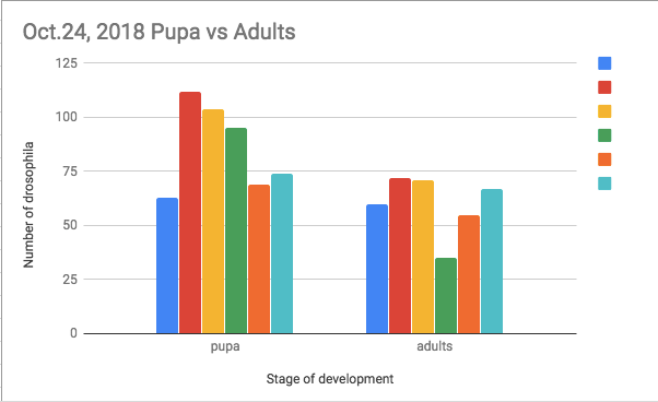
As Raegan and I continue to monitor the progress of our population cross, it is surprising to note that our flies continue to grow at an exponential rate. Using the same key as our last graph(s), it is evident that each tube has experienced significant growth, yet the relative rank regarding populations among tubes has stayed the same. However, whereas on October 17th tubes A, B, and C combined for 239 pupa and flies compared to D, E, and F’s 219, A, B, and C combine for 482 with D,E, and F combining for only 395. Thus, on October 17th the first three tubes represent 109.1% of the second three; this proportion has changed as the first three tubes now represent 122.0% of the second three. Strangely enough, this means that there have probably been not only more flies and pupa in the first three tubes throughout the experiment, but also that the total populations of the first three are increasing at a greater rate despite the fact that there is only one female in tubes A, B, and C in comparison to the three in D, E, and F. Concurring this is the fact that tube A has increased in total population by 339.3%, B by 62.8%, C by 78.6%, D by 46.0%, E by 195.2%, and F by 60.2%. Possible sources of error include the fact that multiple flies escaped during the practice of counting; our food has softened and has buried some flies, pupae, and larvae; and that we are still subtracting two from our population counts to account for parents despite the fact that some tubes had 4 parents and that the parents could have died in multiple tubes.


Eye Color Cross…A Time to Learn

IMG_0464.jpeg

In this first experiment of the year, we set out to recreate Thomas Hunt Morgan’s fly eye color cross. Although we hope to make a formal chart depicting our findings in the future, it is worth noting that our vial labelled “F2” resembled what we hoped the F1 cross would look like and vice versa. Hopefully this is just mis-labeling but could be a product of us isolating the flies too late.

Population Study with Drosophila Progress


Figure 1                                                                                                            Figure 2

Raegan and I have recently collected data pertaining to our Drosophila population cross. The first data, where, in the figures, the dark blue represents tube A, the red represents tube B, the yellow tube C, the green tube D, the orange tube E, and the light blue tube F, we collected on Friday October 12th, 2018 while the second data (with the same legend) we collected on October 17th, 2018. As the figures highlight, all tubes experienced significant population increase, yet there is no significant distinction between tubes that started with one male and one female (A-C) rather than those that started with one male and three females (D-F). I originally thought that a notable distinction would occur between these two subsets, yet our sources of error (i.e. miscounting, escape of flies, and accidental killing of flies while counting) do not seem large enough to have such a significant effect. Thinking about how scientists model population, it can be assumed that, in terms of a logistic growth model, our fly population has grown exponentially as our first data collection represented approximately two weeks after we had isolated the “p” generation whereas our second data collection (where tube B grew by 286.1%, C 445.5%, D 269.7%, E 840%, and F 419%) was only five days after the first. Updates will follow regarding this particular experiment!

Explaining ALS

In typical cases of ALS, motor neurons cells in both the motor cortex of the brain and motor neurons in the spinal cord die, although the disease may, sometimes, extend itself to other regions of the brain. Although the cause of ALS is still, relatively speaking, a mystery, one of the defining features of a patient afflicted by the degenerative disease is the TDP-43 protein in the form of an inclusion body. Inclusion bodies are found to “clog up” the cytoplasm of a neuron. ALS can also be inherited, allowing neurologists to examine the genes responsible for the symptoms in order to determine a cure (and also a sense of the disease’s origin). The genes found to be associated with the disease are observed to be related to “…protein degradation, RNA processing, and the [cell’s] cytoskeleton…” (Wikipedia). Furthermore, Excitotoxicity, a high concentration of calcium in neurons, has been observed to play a role in the death of them; likewise, neurofilament accumulation has been observed in a few cases. Ultimately, with motor neurons dead, the body is not able to convert a stimulus propagated by sensory neurons into an action as the receiver of this signal, the motor neurons, become unresponsive. Furthermore, these neurons are no longer able to carry out a muscular response as their death inhibits them from distributing any electrochemical signal elsewhere.

Screen Shot 2018-09-24 at 8.12.18 AM

For this post, I used https://en.wikipedia.org/wiki/Amyotrophic_lateral_sclerosis, specifically the pathophysiology section as well as Chapter 2 in our textbook Nerve Cells, Neural Circuitry, and Behavior by Eric R. Kandel, Ben A. Barres, and A.J. Hudspeth.

Eyes and Vision

  • Looking at Cajal’s diagram of the retinal cells in an eye alongside the diagram above, I believe that component “a” of the sketch represents the pigmented epithelium. Furthermore, I believe cell type “b” is representative of rods while cell type “c” is indicative of the wider cone. Although I do not know what type “d” or “f” are, structure “e” appears to be the nucleus of the cone. Referring to the diagram on the right once more, “g” appears to be a horizontal nerve cell while “h” seemingly represents a bipolar cell and “i” a ganglion cell. Recalling the reading from “Nerve Cells, Neural Circuitry, and Behavior” by Eric R. Kandel, Ben A. Barres, and A. J. Hudspeth, I would guess the “j” is in some way representative of a mass of neural connections and processes connecting the aforementioned ganglion cells to the optic nerve “m.”
  • Wikipedia describes a photoreceptor as a “neuroepithelial cell” as the various rods and cones that make up our eyes and the eyes of many other organisms communicate light and color to the brain. Photoreceptors are often on the surface of a retina as they represent the inception of the conversion from light into electrochemical signals.
  • As often is the case in biology, the form of photoreceptors meets its function as rhodopsin, a pigment that absorbs  light is stacked much like the thylakoid grana found in the chloroplasts of plant cells. Although most prevalent in rods, rhodopsin works in both of the aforementioned photoreceptors as specific proteins are able to absorb certain wavelengths of light, giving, for instance, the red coloration to a wild-type fly’s compound eye.


Sources: Rhodopsin, the eye and vision powerpoint; https://en.wikipedia.org/wiki/Photoreceptor_cell; “Nerve Cells, Neural Circuitry, and Behavior” by Eric R. Kandel, Ben A. Barres, and A. J. Hudspeth

The Brain and Behavior

According to “The Brain and Behavior” by Eric R. Kandel and A.J. Huspeth, Broca’s area along with Wernicke’s area and the analogous zones of the right hemisphere have distinct processing functions that are interconnected during the process of speech and word comprehension. According to the aforementioned source, Broca’s area (in the frontal lobe) is responsible for the understanding of sentence structure and syntax, the underlying rules dictating the way in which we speak (According to Wernicke’s model of distributed processing, Broca’s area also has some motor functions). In contrast, Wernicke’s area (in the temporal lobe) is related to the understanding of a word’s meaning, not necessarily its emotional connotations, but rather its definitions. In this way, Wernicke’s area is described as receptive rather than expressive (although Broca’s area is not solely used for expression). Keeping this distinction, the analogous structure to Broca’s area in the right hemisphere of the brain is responsible for the expression of emotional nuance in speech with regards to the ways in which words are pronounced and sentences are structured. Furthermore, the analogous structure to Wernicke’s area is responsible for the understanding of various intonations and the multiple ways in which the same word might be expressed to elicit an emotional response. Ultimately, the innate capacity for language is predicated on all of these regions but the distinction of a “natural language” highlights how Broca’s area (and perhaps the analogous area in the right hemisphere) is perhaps most critical. As the brain has developed over time, it is important to keep in mind that it is a series of processing units. As language was initially being developed, certain structures and syntax were most conducive to understanding as they could be most easily processed and replicated. Thus, imbedded within all of us are specific rules of grammar that allow our brain to wrap our head around various languages most easily. (I used “The Brain and Behavior” by Eric R. Kandel and A.J. Hudspeth for this assignment)Screen Shot 2018-09-11 at 9.07.20 PM.pngScreen Shot 2018-09-11 at 9.09.08 PM.png

The Emergence of the Compound Eye


  • Emergence occurs when a system of parts is able to accomplish tasks or is imbued with certain features that are only brought about by the act of the combination of said parts as these components combine their traits and properties in a way that forms a novel body.
  • The complex psychological phenomena we experience on a daily basis regarding our persona and our emotional tendencies that make us feel uniquely human can be distilled, or rather reverse-emerged, down to relatively simple components. In the same way that the majority of rationale and language processing often occurs in very specific sections of the brain (the left hemisphere), “affective elements” (The Brain and behavior, Erik R. Kandel, A.J. Hudspeth) of our psyche cause interconnected sections of our brain to become active and to promote these intangible, often fleeting feelings. Ultimately, neural synapses and brain cells located in certain areas of our brains (for the most part the right hemisphere) and communicating with electrochemical signals control our temperament and thought processes with chemicals and other means with which to disrupt our homeostatic nature.
  • The compound eye of arthropods as we know them today serves as a major clue regarding the development of vision among organisms. In terms of the evolution of such an eye, there are two conflicting schools of thought as to whether the compound eye was originally present in onychophorans (not a true arthropod) or whether the eye evolved later, once the arthropod lineage truly came to be. (en.wikipedia.org) The arthropod eye has similar functional elements with regards to the human eye with cones and lenses while also having novel receptive elements such as the rhabdom (the replacement for rods). (www.britannica.com) The Ommatidium, the eye of flies and other arthropods, also contains various pigment cells that affect eye color (www.ncbi.nlm.nih.gov), aid in perception, and contribute to the overall structure of the eye (the eye actually generates more of these cells than is required, causing some to lyse (www.ncbi.nlm.nih.gov)). (www.sdbonline.org)Much as this eye has similar or analogous features to the human eye, the function is comparable as light is inflected via the lense, passes through various receptors and is shuttled to the brain via optic nerve(s). (en.wikipedia.org) The arthropod eye is one of nature’s most elegant examples of emergence as structural and sensory elements come together to form a system that allows arthropods to detect danger or food in their surroundings. A pigment cell, cone, or lens could accomplish none of these tasks on its own, yet the ocular complex mimics the structure of some of the world’s most perceptive eyes.Screen Shot 2018-09-11 at 7.44.26 AM (www.ncbi.nlm.nih.gov)